Learn how retinal imaging works, what it tests for, and why it's an important part of your eye health.

Has your eye doctor ever taken a picture of your eyes? This is called retinal imaging. It’s a special way for your doctor to get a detailed view of the inner and back surface of your eye, including your retina, the optic nerve, and other important eye structures, according to the Cleveland Clinic.
It’s a very important part of your comprehensive eye exam, because it helps your doctor to spot signs of serious eye problems, including macular degeneration, glaucoma, diabetes-related eye conditions, and more. Here’s what you need to know about this diagnostic tool.
1. Retinal Imaging Helps Detect Eye Problems Early
Retinal imaging is super useful because it helps doctors catch problems early, even before you notice anything wrong with your eyes. Some of the main things it looks for are:
- Diabetic eye problems: If you have diabetes, your eyes can be affected. Retinal imaging checks for early signs of damage. (Learn more about diabetes and your eyes.)
- Glaucoma: Glaucoma is a condition that can damage the optic nerve and cause blindness if not treated. Retinal imaging helps catch it early.
- Macular degeneration: Macular degeneration affects your central vision and can make it hard to see things clearly. Retinal imaging can detect this in early stages.
- Retinal detachment: If your retina starts to pull away from the back of your eye, it can lead to serious problems. Retinal imaging helps your doctor spot this before it gets worse.
If you have been diagnosed with an eye disease, your eye doctor will use retinal imaging to help monitor the progression and determine the best treatment plan for you.
Has it been a while since your last eye exam? Now’s the time to book an appointment!
2. It’s a Part of a Comprehensive Eye Exam
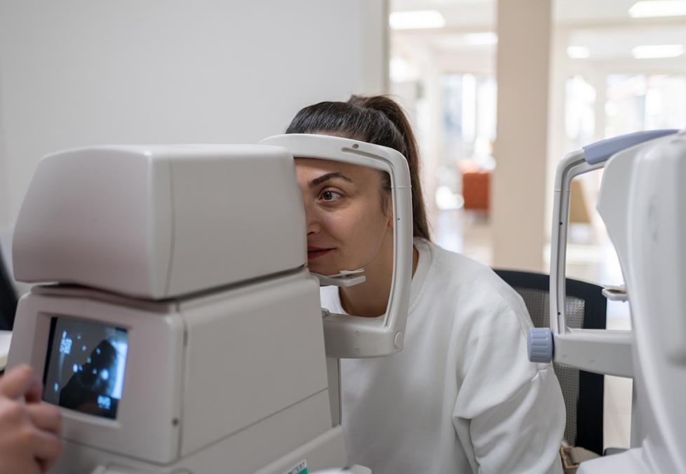
Retinal imaging usually happens as part of a comprehensive eye exam. First, you might take vision tests where you read letters on a chart or look through lenses to check your prescription. After that, you sit at a special machine where the doctor takes a picture of your retina. The best part? It’s quick, painless, and doesn’t involve touching your eye.
3. It is Different Than Eye Dilation
You may be familiar with another way doctors check the retina — eye dilation. This involves using special eye drops to make your pupils (the black part of your eye) larger. This allows the doctor to see more of the inside of your eye. It may be used when they need a closer look. It takes about 20 minutes for the eye drops to widen your pupils, while retinal imaging is immediate. The side effects of dilation include blurry vision that can last for a few hours afterward, unlike retinal imaging.
4. Retinal Imaging Can Detect Problems Beyond the Eyes
Not only does retinal imaging help your eye doctor, but it can also show signs of other health issues. For example, high blood pressure or signs of a stroke can show up in your eyes before they appear anywhere else.

5. There Could Be an Extra Cost
Retinal imaging might not be included in a regular eye exam, so there could be an extra cost. (The price is usually between $20 and $40 in addition to the exam.) Your eye doctor may recommend retinal imaging based on your health history and other personal risk factors for certain eye diseases and conditions.
Some vision insurance plans cover retinal imaging. It’s always a good idea to ask your insurance company about services that are included in your plan before your appointment.
Regular eye exams are the key to keeping your vision clear and your eyes healthy. Make sure to see your eye doctor regularly, and they’ll help you decide the best diagnostic tests for you.
Medically reviewed by: John Bankowski, O.D.
See our sources:
Retinal imaging overview: Cleveland Clinic
Detecting eye diseases: National Eye Institute
Retina and vision: MedlinePlus

